



A biopsy is a medical procedure where a doctor removes cells or tissue from a patient’s body so they can be examined visually. Usually, a microscope does the heavy lifting. The goal is straightforward. Doctors need to see what is happening at a cellular level. This is the standard step for diagnosing both malignant and benign tumors. It also provides diagnostic data for organs like the liver or pancreas.
Getting the sample isn’t always the same. Methods vary based on where the problem is located. Doctors use needles for aspiration. They use swabs. Sometimes they scrape with a curette. Trephining a bone or using forceps to excise tissue with an electric snare are also options. The technique depends on the instrument and the target area.
Preparing the Sample for the Microscope
Once the tissue is collected, it doesn’t just sit there. It has to be prepared. The sample is fixed in ethanol, formalin, or another suitable fixative. This preserves the structure. Next, it is infiltrated with paraffin. This allows it to be sectioned into very thin slices. These slices go onto glass slides.
Before looking under the lens, the tissue undergoes deparaffinizing. Then it is stained. Staining defines the cellular characteristics. A surgical pathologist uses a microscope to examine the tissue. This process leads to a precise diagnosis.
Speed matters in some cases. During surgery, time is critical. Tissue samples can be immediately frozen. Slices are made with a microtome and placed on a slide. They are rapidly stained. The surgeon gets a “frozen section” report within minutes. This allows for immediate decisions in the operating room.
Minimally Invasive Techniques
A needle biopsy is often the simplest route. It is the least-disruptive way to obtain tissue for pathological examination. You have two main choices here. A large cutting needle obtains a “core” of tissue. A small-gauge needle offers a different approach.
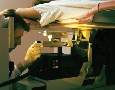
Small-gauge needles perform fine-needle aspiration biopsy. The needle inserts into the area of interest. Suction draws the tissue into the needle. This is common for breast masses. Stereotaxic surgery helps guide fine-needle biopsy. It evaluates breast lesions that aren’t palpable but show up on mammography.
Endometrial biopsy is another form. It targets the uterus specifically. A curette goes into the uterus. Suction collects cells from the internal lining. The specimen contains cells from that internal surface.
Surface Sampling and Cytology
Sometimes you don’t need to cut deep. Abrasion gets cells from surface and subsurface layers of lesions. A brush or spatula does the work. This method works well for epithelial-lined body cavities. Think cervix, vagina, bronchus, or stomach.
These collected cells are examined using the Papanicolaou technique. This is the famous Pap smear. George Nicolas Papanicolaou, a Greek physician, developed the staining and fixing method. The test examines cervical cells fixed and stained on a slide.
The impact of this simple test is huge. Sampling cervicovaginal cells has greatly reduced the incidence of cervical cancer. The technique isn’t limited to the cervix. It applies to cells from other surfaces too.
Exfoliative cytology is an alternative to traditional biopsy. It is quick and simple. Cells shed from body surfaces, like the inside of the mouth, are collected. You examine them directly. This only works for surface cells. It often requires additional cytological analysis to confirm results. It’s a useful tool, but not a standalone solution for everything.
Cutting It Out
An excisional biopsy removes the entire lesion. This is the total removal of the area to be examined. It is most often used for skin lesions. The advantage is clear. The pathologist gets the whole lesion. You minimize the chance that a cancer in part of the lesion would be missed.
In contrast, an incisional biopsy takes only a portion. The whole tumor isn’t removed. This happens when size or location prohibits complete excision. It is also used when a needle biopsy fails. If the needle sample doesn’t provide enough info, an incisional biopsy steps in. The surgeon cuts out a piece big enough to diagnose.
Why do we stick with these methods when technology advances? Because seeing the structure directly still beats guessing. The microscope doesn’t lie about the cellular architecture. It shows the chaos of cancer or the calm of benign tissue.
Some argue that non-invasive scans are replacing biopsies. But scans show shadows. They show density. They don’t show cell shape. You still need the tissue. You still need the stain. The pathologist still needs to look.
The process is mechanical. It is tedious. It requires precision. A needle goes in. A slide comes out. A diagnosis follows. It is the bridge between suspicion and certainty.
And yet, the waiting period for the report can feel like an eternity. You lie there. The needle is gone. The bandage is on. You think about what the cells might say. You don’t know. Nobody knows until the pathologist signs the name.